Bacterial porcine pneumonia
This paper introduces the main pathological changes of bacterial pneumonia. Although most of them are opportunistic, there are two kinds of primary bacteria that can cause pathological changes on their own.
The bacteria that cause porcine pneumonia and can cause pathological changes by themselves can be classified as primary pathogens. Including Mycoplasma pneumoniae, Actinobacillus pleuropneumoniae, Bordetella bronchisepticum and Pasteurella multocida. Secondary pathogens are bacteria that play a role in previously infected primary pathogens or in animals with low immunity; most of them are Pasteurella multocida, Streptococcus suis, Glasiella (formerly known as Haemophilus parasuis) and Cryptobacter pyogenes.
Among the primary pathogens, Mycoplasma hyopneumoniae is the pathogen of epidemic pneumonia, which can cause serious economic losses to pig farms, which is related to the decrease of average daily gain, the increase of feed conversion rate and the increase of mortality caused by secondary infection. This kind of prokaryote belongs to the class Chorioderma and is highly pleomorphic. It is characterized by lack of cell wall, circular double-stranded DNA, polysaccharide capsule with radial fibers, and is difficult to culture.
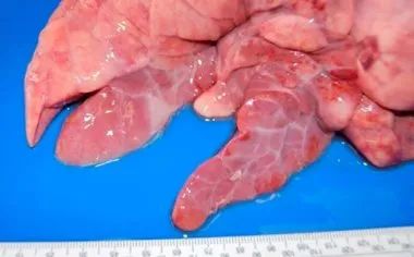
In order to play a pathogenic role, it binds to the cilia of respiratory epithelial cells, resulting in the loss of cilia and changes in the function of the mucociliary system, which means that lung secretions, inhaled particles and pathogens entering the airway cannot be removed. so they move to the lower respiratory tract, complicating the respiratory process. In the lungs, the bacteria can cause bronchial interstitial pneumonia, which is macroscopically characterized by a red consolidation area in the ventral cranial side of the lung, which is clearly defined with other solid areas (figure 1). Under the microscope, the proliferation of lymphoid tissue around bronchioles, bronchioles and blood vessels can be observed, which is characteristic of this process (figure 2).

Figure 1: bronchial interstitial pneumonia caused by Mycoplasma hyopneumoniae
Figure 2: peri-bronchiolar lymphoid tissue hyperplasia caused by Mycoplasma hyopneumoniae.
Streptococcus pleuropneumoniae (APP) is a kind of capsular gram-negative cocci. There are two biotypes: nicotinamide adenine dinucleotide (NAD) dependent biotype I and nicotinamide adenine dinucleotide independent biotype II. There are 18 serotypes: 1-12 and 15-18 belong to biotype Ⅰ, while serotypes 13 and 14 belong to biotype Ⅱ. Streptococcus infectious pleuropneumoniae can produce four toxins, Apx Ⅰ, Ⅱ and Ⅲ specific to some strains, and Apx Ⅳ common to all strains, which is why Apx Ⅳ is the target of several indirect ELISA diagnostic tests. The bacteria colonized in the tonsil, moved down to the lower respiratory system, adhered to alveolar lung cells, where they secreted toxins, produced hemolysis and cytotoxicity, and changed the function of endothelial cells. the phagocytosis of alveolar macrophages will lead to the activation of coagulation cascade reaction, the formation of microthrombus and ischemic necrosis, which are the most characteristic of the pathogen.
The type of pulmonary lesion caused by it is fibrin bronchopneumonia. In hyperacute and acute cases, the lesions mainly occur in the back and distal part of the lung (figure 3), accompanied by coagulative necrosis (infarction) and bleeding, fragile and marble when cut. Microscopically, fibrin and degenerative leukocytes can be seen in the alveolar cavity, bronchioles and bronchioles, accompanied by necrosis and bleeding of varying sizes, alveolar and interstitial edema, and fibrin pleurisy. These same lesions may be caused by some highly virulent strains of Bacillus tumefaciens, so in order to help us make a correct diagnosis, we need to analyze clinical history, macroscopic lesions and microbiology.
Infectious pleuropneumonia can also occur in chronic lesions, acute lesions cause blood-borne transmission, the lungs can see white, different sizes of multifocal lesions.
Figure 3: caudate lobe lesions produced by Actinobacillus pleuropneumoniae
Most Pasteurella multocida strains are secondary pathogens, which can cause lesions ranging from fibrin bronchopneumonia to catarrhal suppurative bronchopneumonia. The former produces fibrin exudate in the alveoli, accompanied by fibrin deposition on the pleural surface. In addition, coagulative necrosis occurs in the lungs due to thrombosis in the blood vessels. In catarrhal suppurative bronchopneumonia, mucous exudates or catarrhal purulent pneumonia are present in the airway (trachea, bronchioles and bronchioles) and alveolar cavity (figure 4). Another cause of this type of bronchopneumonia, especially in the first few weeks of life, may be bronchial septicemia or non-progressive atrophic rhinitis.
Figure 4: suppurative bronchopneumonia: cranio-abdominal consolidation is enlarged, with a small yellow-and-white area (shown by the arrow) corresponding to the alveoli filled with purulent material. Interstitial edema was also observed.
Bacteria such as Streptococcus pyogenes and Streptococcus suis may be associated with embolic pneumonia, umbilical phlebitis, skin or hoof bacterial infections (suppurative streptococcus), or endocarditis (Streptococcus suis), in these cases, bacteria pass through the blood to the lungs from these lesions. Macroscopically, multifocal lesions are distributed throughout the lungs, whitish in color, between 1 mm and 2 cm in diameter, surrounded by reddish circles in the most severe cases, and rapidly evolve into abscesses (figure 5).
Figure 5: embolic pneumonia caused by Pseudomonas aeruginosa.
Figure 6: fibrous pleurisy observed at the slaughterhouse.
Related
- On the eggshell is a badge full of pride. British Poultry Egg Market and Consumer observation
- British study: 72% of Britons are willing to buy native eggs raised by insects
- Guidelines for friendly egg production revised the increase of space in chicken sheds can not be forced to change feathers and lay eggs.
- Risk of delay in customs clearance Australia suspends lobster exports to China
- Pig semen-the Vector of virus Transmission (4)
- Pig semen-the Vector of virus Transmission (3)
- Five common causes of difficult control of classical swine fever in clinic and their countermeasures
- Foot-and-mouth disease is the most effective way to prevent it!
- PED is the number one killer of piglets and has to be guarded against in autumn and winter.
- What is "yellow fat pig"? Have you ever heard the pig collector talk about "yellow fat pig"?


