How to treat pig disease: what are the methods for diagnosing porcine mycoplasma pneumonia?
Porcine mycoplasma pneumonia is the simplest self-replicating prokaryote without cell wall and between bacteria and viruses. Porcine mycoplasma pneumonia is an extracellular pathogen that binds closely to porcine ciliated epithelial cells in the lower respiratory tract rather than invading the body through tissue. During infection in vivo, porcine mycoplasma pneumonia recognizes the specific receptors of ciliated cells in the respiratory tract and propagates in the respiratory tract. It is generally believed that the specific adhesion between porcine mycoplasma pneumonia and respiratory mucosal cilia is a prerequisite for causing mycoplasma pneumonia. In the process of feeding and management, pigs infected with porcine mycoplasma pneumonia alone, pig body weight and feed conversion rate will not decrease, nor will it affect the increase of fat and protein in pigs. It will not affect the growth of pigs in the short term. However, when porcine mycoplasma pneumonia is co-infected with other pathogens, the clinical symptoms of pigs are aggravated.
The methods for diagnosing porcine mycoplasma pneumonia are as follows:
1. Clinical diagnosis of porcine mycoplasma pneumonia.
The main clinical symptoms of porcine mycoplasma pneumonia are chronic dry cough, growth retardation, high morbidity, low mortality and slow spread after infection. However, Mycoplasma hyopneumoniae is easy to be co-infected with other pathogens, such as Actinobacillus pleuropneumoniae, Porcine Reproductive and Respiratory Syndrome virus, Pasteurella pneumoniae and so on, which complicates the clinical symptoms of porcine mycoplasma pneumonia. and some infected pigs do not have clinical symptoms, therefore, through clinical symptoms can only suspect porcine mycoplasma pneumonia.
2. Pathological diagnosis of porcine mycoplasma pneumonia.
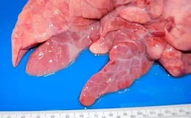
Pathological examination showed that the lesions were mainly in the lungs, hilar lymph nodes and mediastinal lymph nodes, and there were no special lesions in other tissues and organs. The characteristic lesions were pulmonary enlargement with varying degrees of emphysema and edema. Symmetrical fusion bronchopneumonia occurred in the cardiac lobe, apical lobe, intermediate lobe and the anterior and inferior edge of the diaphragmatic lobe in both lungs, in which the cardiac lobe was the most obvious, followed by the apical lobe and the middle lobe.

3. Pathogen detection technique for diagnosis of porcine mycoplasma pneumonia.
1) isolation and culture of pathogen
The isolation of mycoplasma pneumoniae from the diseased site is a specific method for the diagnosis of MPS in pigs. However, porcine mycoplasma pneumonia is one of the most difficult pathogens to be isolated and identified. it grows slowly and is often covered up by the overgrowth of porcine nasal mycoplasma, and it is difficult to isolate porcine mycoplasma pneumonia after drug treatment or rehabilitation. Although the isolation rate of porcine mycoplasma pneumonia is gradually increased by improving the culture medium and isolation method, but in most cases, the diagnosis of isolation and culture is still not feasible.
2) Immunoenzyme technique
Enzyme-linked immunoperoxidase and indirect immunoperoxidase tests are used to detect Mycoplasma pneumoniae in diseased lungs. It overcomes the shortcomings that fluorescent antibody technique can not effectively detect chronic infected pigs, need fresh frozen tissue and fluorescence microscope, and samples can not be preserved for a long time. During ELIP examination of frozen sections of the lung and bronchial smears, mycoplasma was stained with reddish brown pleomorphic particles and the response was stable. Doster et al used formalin fixed paraffin-embedded pig lungs, indirect peroxidase test to detect Mhp, Mhp with clear structure can be observed by optical microscope, and the samples can be preserved for a long time for re-evaluation, which is more sensitive than fluorescent antibody method. This method can be used when there is no fresh tissue or other specific tests.
Summary:
There are many methods for the diagnosis of porcine mycoplasma pneumonia. the above are several commonly used detection techniques reported at home and abroad, each of which has its own advantages and disadvantages. In clinical application, in addition to selecting appropriate detection techniques according to experimental conditions and purposes, comprehensive diagnosis should be made combined with clinical symptoms and histopathological lesions in order to obtain correct and reliable diagnostic results.
Related
- On the eggshell is a badge full of pride. British Poultry Egg Market and Consumer observation
- British study: 72% of Britons are willing to buy native eggs raised by insects
- Guidelines for friendly egg production revised the increase of space in chicken sheds can not be forced to change feathers and lay eggs.
- Risk of delay in customs clearance Australia suspends lobster exports to China
- Pig semen-the Vector of virus Transmission (4)
- Pig semen-the Vector of virus Transmission (3)
- Five common causes of difficult control of classical swine fever in clinic and their countermeasures
- Foot-and-mouth disease is the most effective way to prevent it!
- PED is the number one killer of piglets and has to be guarded against in autumn and winter.
- What is "yellow fat pig"? Have you ever heard the pig collector talk about "yellow fat pig"?


