Research Progress of Porcine Coronavirus: transmissible gastroenteritis virus
Brief introduction
Coronavirus belongs to net nest virus purpose RNA virus, which has two subfamilies:
The family Coronaviridae is composed of α-coronavirus, B-coronavirus and C-coronavirus.
The family Tulongviridae, including the genera Tulong virus and the carp virus.
These viruses can cause five diseases in pigs, in chronological order:
1. Infectious gastroenteritis (TGE-1946)
2. Hemagglutinating encephalomyelitis (HEV-1962)
3. Porcine epidemic diarrhea (PED-1977)
4. Porcine respiratory coronavirus (PRCV-1984)
5. Porcine Delta coronavirus (PRCV-1984)
Three kinds of porcine coronavirus are associated with digestive diseases (TGE, PED and PDCoV). Porcine respiratory coronavirus (PRCV) is associated with respiratory diseases. Hemagglutinating encephalomyelitis virus can cause vomiting and emaciation, and encephalomyelitis.
There have been no reports of human infection with porcine coronavirus.
1 transmissible gastroenteritis virus
Transmissible gastroenteritis virus is a coronavirus, which has been rarely found since the late 20th century. It was first reported in 1946 that it reached its highest prevalence in Europe and the United States in the 1970s, 1980s and 1990s, and more than 95% of European pig farms were positive in the 1980s. It is a highly contagious virus that can cause diarrhea, dehydration, occasional vomiting and high mortality in piglets. Porcine transmissible gastroenteritis virus has been completely sequenced and only one serotype is known.
There is homology between porcine transmissible gastroenteritis virus and bovine and human coronavirus and porcine respiratory coronavirus. From an epidemiological point of view, there are both epidemic and endemic cases. It is a relatively heat-resistant virus that is resistant to low pH values and many disinfectants. Inactivated in less than two hours at 37 ℃. It is very sensitive to light and extremely resistant to freezing. The virus can survive for a long time in frozen corpses. This means that most disease outbreaks occur in cold months (winter).
pathogenesis
The route of transmission is fecal-oral transmission. The virus propagated mainly in gastrointestinal cells. 4-5 hours later, it was found that the virus existed in the cytoplasm of infected cells, mainly in the bottom of small intestinal villi. It causes villus atrophy, which significantly reduces nutrient absorption and leads to osmotic diarrhea, while reduced sodium and glucose transport worsens osmotic diarrhea, leading to hypoglycemia in affected animals. At the same time, it proliferates in different extra-intestinal organs such as lungs (alveolar macrophages), breast tissues and lymph nodes.
IgA in colostrum can protect 6-12-week-old piglets. The active antibody appeared one week after infection, which lasted for 6 months in fattening pigs and two years in sows. After the disappearance of clinical symptoms, the virus can persist in pigs, and it can be detoxified through feces within 10 weeks, thus becoming an asymptomatic carrier with the risk of spreading infection. Breeding sows can spread the virus through breast milk. Other routes of transmission include trucks, boots, dung, and even dogs and cats, because the virus stays in their digestive tract for two weeks and can be excreted. The incubation period was only 1-2 days, and the duration of clinical symptoms was 7-10 days.
Clinical symptoms
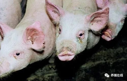
In pig farms without transmissible gastroenteritis, the most typical infections are acute infections and episodes of acute cases, showing sudden and severe diarrhoea, affecting all stages of production within a few days. Diarrhea is severe, watery, yellowish green, and sometimes smelly.
In suckling piglets, vomiting is common, accompanied by loss of appetite, no fever or neurological symptoms. Dehydration occurs within 24-48 hours and the mortality is high. The mortality rate of piglets less than 1 week old can be as high as 100%, 50% in the second week of lactation and 25% in the third week. The piglets died after dehydration, stomach dilatation, breast milk and hemorrhagic stasis spots in the small intestine, hypertrophy of mesenteric nodules (characteristic lesions), intestinal microvilli atrophy (duodenum, jejunum and ileum), intestinal wall thinning, jejunal epithelial cell necrosis and enzyme activity decreased.
Mixed infection of gastrointestinal bacteria such as Escherichia coli and Clostridium is common in piglets, which worsens and prolongs clinical symptoms. Pigs over one month old have similar symptoms, but much lower severity, high morbidity and extremely low mortality.
In pig farms where the disease is endemic, pigs are infected later until they enter the fattening stage. The main effects on fattening pigs are slow growth and low feed conversion rate.
In breeding sows, except for diarrhea, the abortion rate and sow mortality increased only slightly within 1-3 weeks. Lactating sows develop anorexia and occasional anorexia.
Diagnosis
The clinical symptoms and pathological changes are quite obvious, but we must differentiate the strains of enterotoxin Escherichia coli, Clostridium, rotavirus, coccidia and Cryptosporidium. The age and clinical symptoms of infected pigs will also help us narrow the diagnosis to coronavirus.
Laboratory analysis confirms that it depends on:
Specific monoclonal antibodies in pig serum were used for ELISA to detect antigens in feces and intestinal contents.
Electron microscopic examination to detect viruses in intestinal contents.
Serum neutralization test was performed 7-8 days after infection and 18 months after infection.
Immunoperoxidase in intestinal tissue was fixed to detect virus in piglets.
The virus was isolated from intestines, tonsils, lymph nodes and feces by PCR.
Treatment
It can only be treated symptomatically. There is no commercial vaccine available. There used to be one in the United States, but it is no longer on the market. Hydration is the main treatment measure, the control of concurrent infection can help us to reduce the impact, while strengthening biosafety measures, prolonging empty column time and strictly controlling environmental conditions are very important for piglets. The main aim is to infect all animals in pig farms as soon as possible in order to obtain active immunity. The main control measures are prevention:
1. Biosafety: used only for clothing, visitor control, feed transportation and animals in pig farms.
two。 Strictly implement all-in-all-out in different production areas. Wash and disinfect with hot water of 65 °C.
3. Prevent cats and dogs from entering the pig farm.
4. Effective disinfection, deratization and pest control procedures.
5. Negative reserve sows and boars enter the breeding house.
6. The reserve sows were isolated and domesticated for 8-9 weeks, and their gastrointestinal symptoms were strictly observed.
7. Drinking water disinfection procedure.
8. Piglets were fed colostrum within 24 hours after birth.
Related
- On the eggshell is a badge full of pride. British Poultry Egg Market and Consumer observation
- British study: 72% of Britons are willing to buy native eggs raised by insects
- Guidelines for friendly egg production revised the increase of space in chicken sheds can not be forced to change feathers and lay eggs.
- Risk of delay in customs clearance Australia suspends lobster exports to China
- Pig semen-the Vector of virus Transmission (4)
- Pig semen-the Vector of virus Transmission (3)
- Five common causes of difficult control of classical swine fever in clinic and their countermeasures
- Foot-and-mouth disease is the most effective way to prevent it!
- PED is the number one killer of piglets and has to be guarded against in autumn and winter.
- What is "yellow fat pig"? Have you ever heard the pig collector talk about "yellow fat pig"?


