Diagnosis and treatment of porcine mold feed poisoning complicated with eperythrozoonosis
1 introduction of the disease
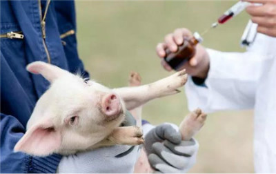
Liu has many years of breeding experience, usually with his own ingredients. At present, there are 30 pigs, including 1 pregnant sow and 1 fattening pig (about 100kg), and the other 28 piglets were purchased from the market one month ago. The piglets developed diarrhea symptoms a few days after they arrived home, and one piglet died at that time, and the condition was controlled by medication. Then immunized with classical swine fever, swine erysipelas, swine lung disease. But within a few days, this batch of piglets fell ill one after another and showed diarrhea symptoms. Liu bought medicine by himself, injected intramuscularly with "Haida" injection, dexamethasone, an sodium coffee, dysentery net, and norfloxacin drinking water, which played a certain role after using norfloxacin water, but still could not control the disease, and two piglets died. Sows and fattening pigs are basically normal.
2 clinical symptoms
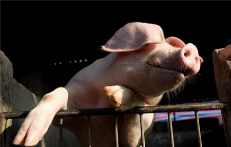
The sick pig is depressed in spirit, loss of appetite or even abolition, black and thin stool, elevated body temperature, shortness of breath, some can not afford to lie down, visible mucous membrane flushing, yellow staining. The skin of the ear, the inside of the limbs and abdomen is slightly purple, finger pressure fades, abdominal pain, some skin is pale, some skin is slightly red, the coat is rough, the ear is cyanotic, the edge is rolled up, emaciated, and the growth is slow.
3 pathological changes
According to the netizens of Pig e-net, it was learned that the main pathological changes of 2 dead pigs were subcutaneous bleeding and subcutaneous fat yellow staining. Oral mucous membrane bleeding, dehydration, laryngeal epiglottic cartilage bleeding, yellow staining, tracheal mucus, tracheal bleeding, a small amount of yellow fluid in the chest, a small amount of yellow fluid in the pericardium, bleeding spots on the surface of the heart, pulmonary hyperemia, edema, and necrotic foci. The spleen is enlarged and softened, and there are many small infarcts at the edge. The liver has many white necrotic foci of different sizes. The liver is hard and brittle. Gallbladder atrophy, less bile, light color, gallbladder mucosal bleeding, inner wall edema. Gastric mucosa at the bottom of the serious congestion, bleeding, there are many ulcers, necrotic spots, mucous membrane easy to fall off, duodenal bleeding, cecal mucosal bleeding, edema, rectal mucosal bleeding, edema, mesenteric lymph node congestion. There are bleeding spots on the surface of the kidney, appearance edema, section valgus, bladder mucosa, slight bleeding, yellow and sticky oily urine. The blood is thin, the color is light, and the blood coagulation is poor.
4 diagnosis
4.1 Laboratory diagnosis
4.1.1 the mucus in the pig trachea was placed on the slide and 1 drop of 20% potassium hydrochloride solution was added to the slide. after the cover slide, a number of radial hyphae and a large number of mold spores could be seen.
4.1.2 take the blood of diseased pigs and dead pigs.
4.1.2.1 smear staining: blood smear, Wright's staining, microscopic examination showed that red blood cells were light pink, there were spherical, circular and other light purplish red particles in red blood cells.
4.1.2.2 Direct microscopic examination: dilute the blood with 2 times the amount of normal saline, drop 1 drop on the slide and cover the slide. Under microscope, red blood cells are serrated, cauliflower-shaped, star-shaped, globular and punctate pathogens can be seen in plasma and erythrocytes, and a cluster of spherical bright pathogens can be seen in erythrocytes.

- Prev
Don't be careless in feeding pigs, eight common symptoms of pig poisoning
Don't be careless in feeding pigs, eight common symptoms of pig poisoning
- Next

What about swine mold poisoning? Prevention and control of mycotoxin poisoning
What about swine mold poisoning? Prevention and control of mycotoxin poisoning
Related
- On the eggshell is a badge full of pride. British Poultry Egg Market and Consumer observation
- British study: 72% of Britons are willing to buy native eggs raised by insects
- Guidelines for friendly egg production revised the increase of space in chicken sheds can not be forced to change feathers and lay eggs.
- Risk of delay in customs clearance Australia suspends lobster exports to China
- Pig semen-the Vector of virus Transmission (4)
- Pig semen-the Vector of virus Transmission (3)
- Five common causes of difficult control of classical swine fever in clinic and their countermeasures
- Foot-and-mouth disease is the most effective way to prevent it!
- PED is the number one killer of piglets and has to be guarded against in autumn and winter.
- What is "yellow fat pig"? Have you ever heard the pig collector talk about "yellow fat pig"?